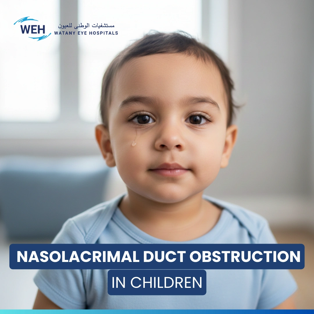
Introduction to Nasolacrimal Duct Obstruction in Children
Nasolacrimal duct obstruction in children (NLDO), also known as tear duct obstruction, is one of the most common eye conditions in infancy. It occurs when tears cannot properly drain from the eye into the nose due to a blocked tear duct. This leads to excessive tearing, mucus discharge, and crusting around the eyes, particularly after sleep.
In most cases, the blockage is congenital, resulting from incomplete opening of the tear duct at birth. Fortunately, many children experience spontaneous improvement or respond well to simple non-surgical treatments.
Early diagnosis by a pediatric ophthalmologist is essential to prevent complications such as chronic dacryocystitis or recurrent eye infections.
What Is Nasolacrimal Duct Obstruction in Children?
Nasolacrimal duct obstruction in children occurs when the passage that drains tears from the eye into the nose becomes blocked or narrowed, preventing normal tear flow. As a result, tears accumulate on the surface of the eye, leading to persistent watering, mucus discharge, and sometimes eye infections.
Normally, tears travel from the lacrimal glands through small openings called puncta, then into the nasolacrimal duct which empties into the nasal cavity. When this pathway is blocked, it causes excessive tearing and may lead to chronic dacryocystitis (infection of the tear sac).
The condition may be congenital (present at birth) due to incomplete duct development or acquired later in life due to infections or trauma. Although often mild, timely diagnosis and management are crucial to prevent long-term complications.
Causes of Nasolacrimal Duct Obstruction in Children
Nasolacrimal duct obstruction in children can result from various congenital or acquired factors. Understanding the underlying cause helps ophthalmologists determine whether the case can resolve spontaneously or requires surgical management.
Common Causes:
- Congenital blockage (Congenital nasolacrimal duct obstruction):
The most frequent cause, where a thin membrane remains at the nasal end of the duct, preventing normal tear drainage.
- Narrow tear duct:
Due to incomplete development of the tear drainage system during fetal growth.
- Recurrent eye infections:
Inflammation or scarring within the duct may block tear flow.
- Facial or nasal trauma:
Injury or fractures near the nose can compress or damage the tear drainage pathway.
- Inferior turbinate hypertrophy:
Swelling inside the nose may physically block the duct’s outlet.
Note: Over 90% of congenital tear duct blockages resolve naturally within the first year of life, making regular monitoring essential before considering surgery.
Check Your Vision Today
Get in touch with us through our hotline or WhatsApp
Hotline
16112Symptoms of Nasolacrimal Duct Obstruction in Children
The symptoms of tear duct obstruction in infants often appear within the first few weeks of life.
Recognizing these signs early helps prevent eye infections and ensures timely treatment.
Common Symptoms:
- Excessive tearing:
Continuous tears on the cheeks even when the baby is not crying.
- Sticky or yellow discharge:
Caused by trapped tears and bacterial growth inside the tear sac.
- Swelling near the inner corner of the eye:
Indicates possible inflammation of the lacrimal sac.
- Redness around the eye:
A common sign of irritation or infection due to the blockage.
- Tears reappearing when pressing near the inner corner:
Confirms obstruction in the nasolacrimal duct.
Tip for parents: Avoid home remedies or excessive cleaning. Always consult a pediatric ophthalmologist for proper diagnosis and treatment.
Diagnosis of Tear Duct Obstruction in Children
The diagnosis of tear duct obstruction in infants is a crucial step to determine the cause of continuous tearing and to differentiate between a simple blockage and other conditions like eye infections or inflammation.
Diagnosis usually involves clinical examination and simple diagnostic tests performed by a pediatric ophthalmologist.
Main Diagnostic Methods:
- Clinical examination:
The doctor checks for swelling of the lacrimal sac, redness, or discharge in the inner corner of the eye.
- Tear flow or dye disappearance test:
A safe dye is placed in the eye to track tear drainage. Delayed clearance indicates blocked tear ducts.
- Lacrimal sac pressure test:
Gentle pressure on the inner corner may cause discharge from the eye, confirming an obstruction.
- Imaging tests (Dacryocystography):
In complex cases, imaging helps identify the exact location of the blockage in the tear duct system.
Professional Tip:
Accurate diagnosis should always be made by a pediatric ophthalmologist, as early detection prevents complications and infections.
Check Your Vision Today
Get in touch with us through our hotline or WhatsApp
Hotline
16112Treatment and Management of Tear Duct Obstruction in Children
The treatment of blocked tear ducts in infants depends on the child’s age and the severity of the obstruction.
In most cases, the blockage resolves naturally within the first few months, but some children may require medical or minor surgical intervention.
Stage 1: Conservative Management (Birth to 12 Months)
This is the most critical stage, as approximately 90% of cases resolve on their own during the first year. Management includes:
-
Lacrimal Sac Massage (Crigler Massage):
This is the primary initial treatment for infants under one year of age.
Regular massage helps open the tear duct and improve drainage. Parents are taught the correct technique by the pediatric ophthalmologist.
-
Home Care:
Gently clean away any discharge using a soft cotton ball dampened with warm water. Avoid directly touching the eye to prevent infection.
-
Medical Treatment:
If a secondary infection is present, a doctor will prescribe antibiotic eye drops or ointments to reduce discharge and prevent the spread of infection.
Stage 2: Minimal Intervention (If Obstruction Persists Beyond 12 Months)
If conservative methods are unsuccessful, the risk of recurrent infections increases, intervention becomes necessary. The most common and effective options are:
-
Probing:
If the duct remains blocked after the child's first birthday, the doctor may recommend probing. This procedure, performed under light anesthesia, involves passing a fine instrument through the tear duct to open the obstruction and restore normal tear drainage.
-
Balloon Dilation:
This procedure uses a very tiny balloon catheter that is inserted into the blocked area of the duct. The balloon is then gently inflated to widen the passage and improve tear drainage.
-
Silicone Stent Intubation:
In some cases, a thin silicone tube is temporarily placed inside the tear duct. This stent helps keep the duct open during the healing process and prevents the obstruction from recurring.
Stage 3: Surgical Options (For Complex or Advanced Cases)
When all previous techniques have failed, or in older children, a final surgical option is considered:
-
Dacryocystorhinostomy (DCR):
This is the definitive surgery for persistent cases. The procedure involves creating a new drainage pathway for tears that completely bypasses the blocked nasolacrimal duct, connecting the lacrimal sac directly to the nasal cavity. DCR is a precise procedure typically performed on children over the age of four.
Possible Complications of Untreated Tear Duct Obstruction in Children
Although tear duct obstruction in infants might seem minor, if left untreated, it can cause serious and painful complications that affect both the eye and the child’s comfort.
Recognizing these risks emphasizes the importance of early diagnosis and proper medical follow-up.
Main complications:
- Recurrent dacryocystitis (infection of the tear sac):
Stagnant tears allow bacteria to grow, leading to redness, swelling, and pain near the inner corner of the eye. In severe cases, it can progress to a tear sac abscess. - Spread of infection (Periorbital cellulitis):
The infection may extend to surrounding tissues, causing eyelid swelling and fever, requiring urgent medical care. - Corneal irritation and visual disturbance:
Chronic tearing and discharge can irritate the cornea, leading to blurred vision or persistent eye discomfort. - Misdiagnosis or delayed treatment:
It can sometimes be mistaken for allergic conjunctivitis or chronic eye infections, delaying proper management.
Watany Eye Hospitals’ advice:
Your child’s eyes deserve early care, timely diagnosis and proper treatment ensure comfort, clear vision, and healthy eyes.
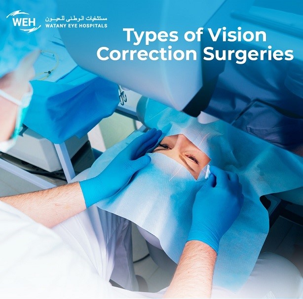
 Blepharoplasty and its Types